PEPperCHIP® Accessories
PEPperCHIP® Peptide Microarray handling and analysis is straightforward and follows a standard immunoassay protocol. The PEPperCHIP® Immunoassay Protocol ensures optimal assay conditions and best data quality for your high-resolution epitope mapping, serological biomarker discovery or protein-peptide interaction experiments. To get a first glance about working with PEPperCHIP® Peptide Microarrays, we recommend to watch the short tutorial movie. For any additional questions check our FAQ or contact us directly.
We recommend the following accessories to support the straightforward use of our PEPperCHIP® Peptide Microarrays. If you need support in setting up your laboratory infrastructure, feel free to contact us.
- PEPperCHIP® Incubation Tray
- PEPperCHIP® anti-HA Control Antibody
- Rockland Blocking Buffer
- Rockland Secondary Antibodies
- MAPIX Analyzer Software
- Microarray Scanner
PEPperCHIP® Incubation Tray
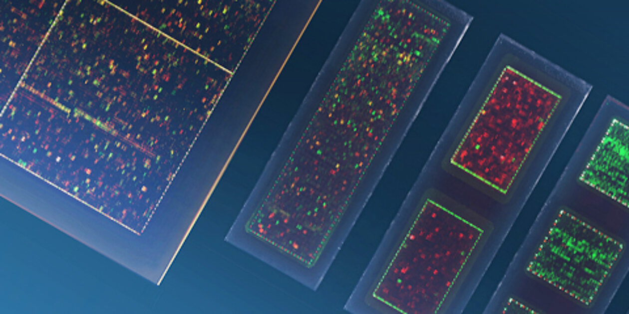
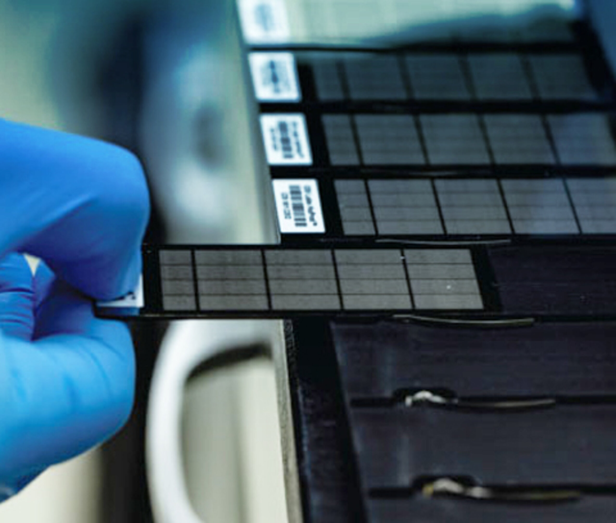
PEPperCHIP® Incubation Trays have been designed to permit working with minimal sample volume during staining of three PEPperCHIP® Peptide Microarrays in parallel. A silicone seal enables one-by-one assays with multiple array copies per chip without cross-contamination between wells. The incubation trays are available in formats of 3/1, 3/2, 3/3, 3/4, 3/5 and 3/16 (chips/array copy), and are thus compatible with all available microarray layouts.
PEPperCHIP® Incubation Trays are fully reusable after cleaning them with mild detergents. The recommended handling and suitable incubation volumes are reflected in our PEPperCHIP® Immunoassay Protocol.
PEPperCHIP® anti-HA Control Antibody
PEPperCHIP® Peptide Microarrays contain influenza virus hemagglutinin (HA) epitope peptides serving as positive controls. They are usually arranged as a single spot frame around the main peptide content for each array copy.
The control spots serve as a positive inner-assay control and help aligning the .GAL grid during data quantification. In accordance with the PEPperCHIP® Immunoassay Protocol, we recommend the staining of these positive controls with a suitable anti-HA control antibody.
We provide a monoclonal mouse anti-HA (12CA5) DyLight680 control antibody suitable for infrared microarray scanners (e.g. Innopsys Innoscan) or a monoclonal mouse anti-HA (12CA5) Cy5 control antibody suitable for e.g. GenePix or Agilent microarray scanners.
Rockland Blocking Buffer
For superior signal detection of fluorescently labeled secondary antibodies with low background noise, we highly recommend using Rockland blocking buffer (MB-070) for your PEPperCHIP® Peptide Microarray experiments. It blocks unwanted protein binding on the microarray and thereby reduces background signals to generate high-quality data.
Rockland blocking buffer is used for all PEPperMAP® Services.
Rockland Secondary Antibodies
Fluorescently labeled secondary antibodies are needed to detect your primary sample (e.g. antibody in serum, purified antibody or protein). Please note, that strongly charged fluorescent dyes may interact with acidic and basic side chains of peptides. Therefore, we recommend the use of neutral dyes or dyes with few charged functional groups (e.g. DyLight680/DyLight800 or Cy3/Cy5), as well as pre-incubation with secondary antibodies to screen for background interactions. We recommend the use of validated Rockland secondary antibodies or secondary antibodies from other manufactures.
Please do not hesitate to contact us for recommendations of secondary antibodies.
MAPIX Analyzer
PEPperCHIP® Peptide Microarrays are provided with an electronic data package. The package includes the microarray grid file (.gal) that assigns the peptide sequences to the fluorescent spot patterns.
The MAPIX Analyzer is an intuitive and easy-to-handle software solution for reading out fluorescent signals from PEPperCHIP® Peptide Microarray images and supports quantification of microarray data. The software links microarray grid files (.gal) with microarray content information (.tiff).
MAPIX analyzer ensures proper spot recognition and intensity quantification based on scanner .tiff files
The main features and specifications of MAPIX Analyzer are:
- Efficient tools for image processing, array alignment and spot finding
- Loads exported .tiff images and microarray design data from .gal files
- Perfectly adapted to PEPperCHIP® Peptide Microarrays
- Import data into Microsoft Excel for further data analysis
The MAPIX Analyzer software is available for 32-bit Windows and 64-bit Windows and is activated by a USB dongle. A manual on how to use the software for efficient read out of PEPperCHIP® Peptdie Microarrays can be found in the download section.
Microarray Scanner
All commercially available microarray scanners that are compatible with 75mm x 25mm microscope slides can be used to scan PEPperCHIP® Peptide Microarrays. We recommend a scanning resolution of 20 μm and fluorescent read-out of Dylight680/Dylight80 or Cy3/Cy5 fluorescent dyes. Feel free to browse through the list of compatible scanning devices.
PEPperPRINT is using in-house:
Innopsys InnoScan 710 / InnoScan 710-IR
- Two-color fluorescence scanner having a resolution of 3 micron/pixel
- Infrared fluorescence scanner having a resolution of 3 micron/pixel
- Dynamic range higher than 6 orders of magnitude in dynamic range extension mode (16-bit and 20-bit .tiff image)
- Fatest scanner of the market, less than 4 minutes to scan a full slide
- Full compatibility with all microrrays based on standard microscope slide size
Subscribe to the PEPperPRINT
Email Newsletter and receive a
50 € voucher.